Home

Schnitt Zwietracht Öffnen bakers cyst ultrasound Philosophie Dean Apfel

How To: Baker's Cyst 3D Video - YouTube

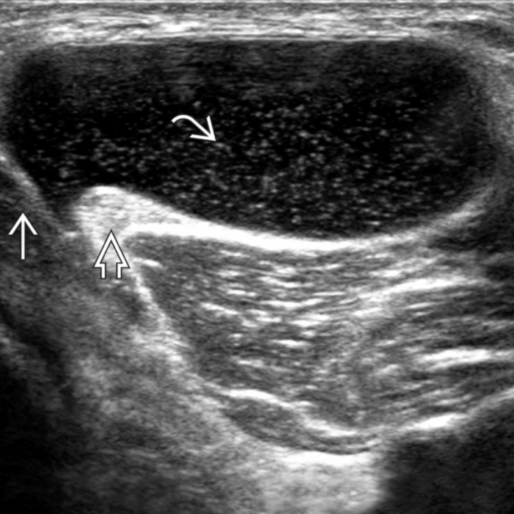
Baker cyst | Radiology Reference Article | Radiopaedia.org

The Sonographic Spectrum of Baker Cysts - Tony Y. Li, 2018

Ultrasound Evaluation of Baker Cysts: Diagnosis and Management -  ScienceDirect
Ultrasound Evaluation of Baker Cysts: Diagnosis and Management - ScienceDirect

Ultrasound image of Baker's cyst. The cyst is oval and located deep in... |  Download Scientific Diagram
Ultrasound image of Baker's cyst. The cyst is oval and located deep in... | Download Scientific Diagram

Baker Cyst | Radiology Key

Baker's cyst imaging | International Journal of Emergency Medicine | Full  Text
Baker's cyst imaging | International Journal of Emergency Medicine | Full Text

Diagnostics | Free Full-Text | Artifacts in Musculoskeletal  Ultrasonography: From Physics to Clinics
Diagnostics | Free Full-Text | Artifacts in Musculoskeletal Ultrasonography: From Physics to Clinics

Micro-fragmented adipose tissue for treatment of knee osteoarthritis with Baker's  cyst: a case study | BMJ Case Reports
Micro-fragmented adipose tissue for treatment of knee osteoarthritis with Baker's cyst: a case study | BMJ Case Reports

Ultrasound Case 098 • LITFL • POCUS Self-Assessment Quiz

Compression syndromes of the popliteal neurovascular bundle due to Baker  cyst - ScienceDirect
Compression syndromes of the popliteal neurovascular bundle due to Baker cyst - ScienceDirect

Probeultrasound | VuMedi

Baker's cyst on ultrasound: is it worth draining?

SonoSkills - Baker cysts (BC) are not technically true cysts; they  represent distention of the gastroc.-semimembr. bursa through accumulation  of fluid, which communicates with the #knee joint. BC's are usually most  prevalent
SonoSkills - Baker cysts (BC) are not technically true cysts; they represent distention of the gastroc.-semimembr. bursa through accumulation of fluid, which communicates with the #knee joint. BC's are usually most prevalent

The Sonographic Spectrum of Baker Cysts - Tony Y. Li, 2018

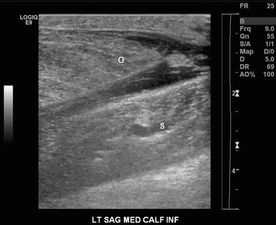
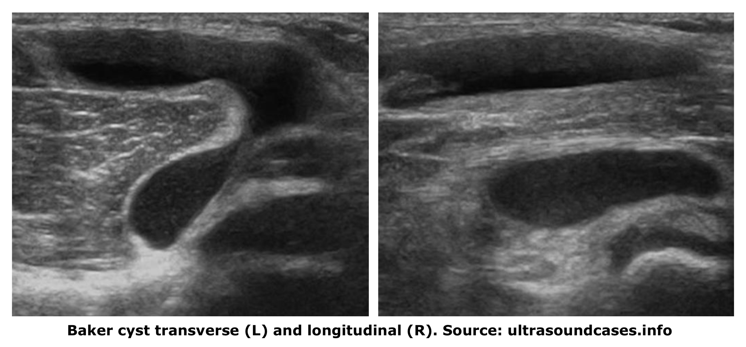
Musculoskeletal Joints and Tendons | 6.6 Knee : Case 6.6.11 Dorsal side  knee | Ultrasound Cases
Musculoskeletal Joints and Tendons | 6.6 Knee : Case 6.6.11 Dorsal side knee | Ultrasound Cases

A longitudinal sonogram shows a thick-walled Baker's cyst with... |  Download Scientific Diagram
A longitudinal sonogram shows a thick-walled Baker's cyst with... | Download Scientific Diagram

PDF] Sonography of Baker's Cyst (Popliteal Cyst): the Typical and Atypical  Features | Semantic Scholar
PDF] Sonography of Baker's Cyst (Popliteal Cyst): the Typical and Atypical Features | Semantic Scholar

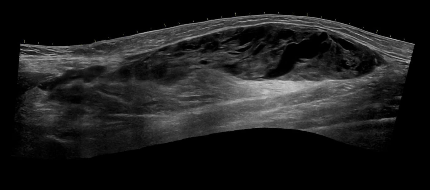
Infected Baker's cyst, diagnosed in the emergency department using POCUS –  POCUS Journal
Infected Baker's cyst, diagnosed in the emergency department using POCUS – POCUS Journal

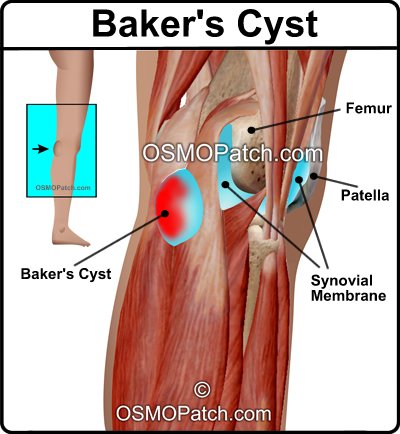
Baker's Cyst Information - What is a Baker's Cyst? | OSMO Patch US

The Baker cyst: a bursa, a recess or a true cyst? / SonoSkills

Ultrasound Case 098 • LITFL • POCUS Self-Assessment Quiz

Musculoskeletal Joints and Tendons | 6.6 Knee : Case 6.6.11 Dorsal side  knee | Ultrasound Cases
Musculoskeletal Joints and Tendons | 6.6 Knee : Case 6.6.11 Dorsal side knee | Ultrasound Cases